In Vivo Molecular Imaging and Therapy Lab, MIT
- Neurobioimaging -
02 Research
In vivo molecular imaging and
therapy is a cutting-edge science technology that evaluates various biological
phenomenon occurring at the molecular or cellular level based on molecular
biology techniques in living subjects using image probes or reporter genes.
This molecular imaging technology is a new technology field that facilitates
translation research between basic and clinical area as a role of connecting
bridge. This imaging area can provide diagnostic and therapeutic information
such as early diagnosis of disease, monitoring of gene expression, and
evaluation of therapeutic response to the drug. We are doing the cutting-edge
researches as the following topics.
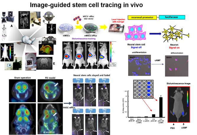
A. Research of image-guided biomedical application in regenerative medicine
Our aim is to understand in vivo characteristics and behaviors of grafted stem cells in a variety of diseases such as their localization, survival, proliferation, and differentiation), which could help researcher to determine optimal cell number, better understand the whereabouts of grafted stem cells and therapeutic mechanism for regenerative therapy.
In addition, this techniques can give useful information in tissue engineering area, showing the supportive effects of various biocompatible scaffolds by imaging the stem cells incorporated within scaffolds of interest such as sponge type, gel-type, and nanofiber-type scaffolds.

<In vivo monitoring of grafted stem cell survival, proliferation, and differentiation based on molecular imaging technology>
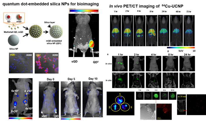
B. Application of cancer-targeted therapy using nanomaterials in nanomedicine area
Based on biocompatible nanomaterials (exosomes, graphene, nanogels, etc.), we have conducted targeted imaging and therapy for the tumor. Image-guided nanotechnology have received great attention until now on, because it can give significant knowledges capable of simply tracing the location of nanomaterials, distribution, and excretion from the body. Research also includes therapeutic radioisotope-based cancer targeting study to maximize the effectiveness of cancer therapy.

<Development of highly sensitive and liver-evading nanomaterials for efficient cancer targeting>
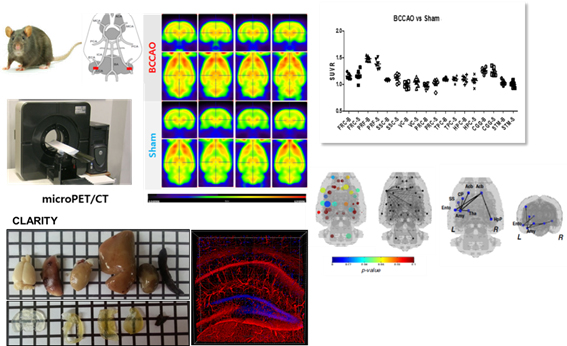
C. Brain imaging / network for diagnosis and therapy of brain diseases
Based on multi-brain molecular imaging technologies (nuclear medicine, magnetic resonance imaging, optical images, etc.), we aim to figure out classification of brain disease, early diagnosis, prediction of prognosis, and evaluation of efficacy of medication through brain circuit analysis of multi-scale units (macro, meso and micro). Currently, we are conducting brain circuit analysis research on various brain diseases (dementia, Parkinson's disease, brain development disorder) using cutting-edge brain technologies such as functional connectivity and brain tissue-clearing CLARITY technique.

<Macro-micro brain imaging and diagnosis of brain disease based on brain network>
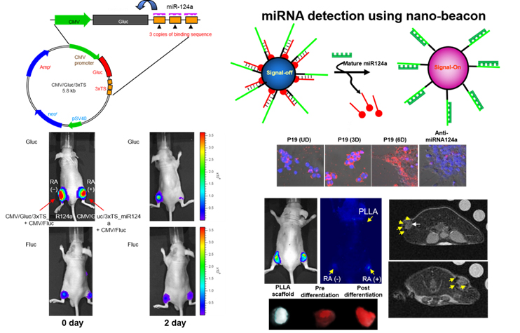
D. RNA imaging techniques for disease early diagnosis
We have assessed the amount of RNA of interest, especially non-coding RNAs by adopting fluorescence and bioluminescence image techniques such as reporter genes or nanomaterial-based molecular beacon targeting disease-specific non-coding RNAs (microRNAs, long noncoding RNAs). These studies help to understand early pathological processes by directly observing non-coding RNAs altered in early disease status. In addition, we are developing diagnostic nano-sensors by introducing sensitive fluorescence imaging technology to detect disease-related RNA.

< RNA expression biomedical imaging studies >
